
Medical specialties
Point-of-Care Ultrasound Diploma
Register interest
An evidence-led diploma in point-of-care ultrasound (POCUS): what a focused bedside scan of the heart, lungs, abdomen and vessels can and cannot answer, how to interpret the images, and how to act on them safely in emergency, acute and critical care. It teaches knowledge, image interpretation and decisions: scanning technique and every ultrasound-guided procedure (vascular access, pleural and abdominal drainage, nerve blocks) need supervised hands-on practice and sign-off elsewhere, and the diploma does not by itself confer accreditation, such as the UK's Focused Ultrasound in Intensive Care (FUSIC) pathway.
Twenty-five weekly sessions across five modules take you from physics, safety and governance, through the heart and circulation, the lungs and pleura, the abdomen, trauma and early pregnancy, to ultrasound-guided procedures and building a service, taught live by practising specialists you can question.
- Course director
- To be announced
- Format
- 25 live weekly sessions on Zoom, in 5 modules
- Fee
- £1,200 (scholarships available)
- Dates
- Confirmed when enrolment opens
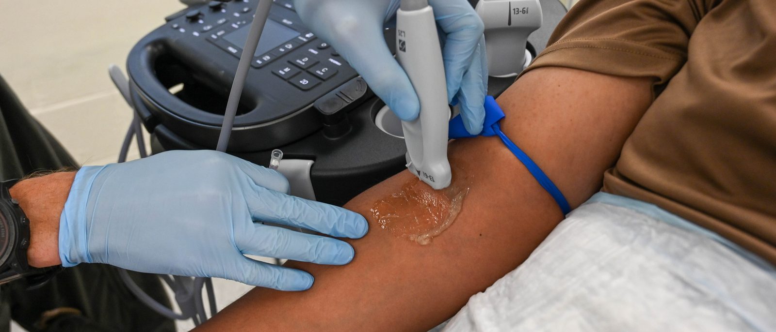
Who it is for
Senior trainees, fellows and early-career consultants in emergency medicine, acute medicine, intensive care and anaesthesia, and other clinicians who use or are learning bedside ultrasound, worldwide.
The programme
25 one-hour sessions in 5 modules, each with its learning objectives. This is the draft programme: the course director reviews and refines it before enrolment opens, so session titles may change.
Module 1 · Sessions 1 to 5Foundations, safety and governance
1. Physics, knobology and image optimisation
- Select the probe and preset for each core application
- Optimise depth, gain and focus to answer a focused question
- Distinguish diagnostic artefacts from misleading ones
- Apply the thermal and mechanical indices to keep scanning safe
2. Training, accreditation and scope of practice
- Describe the UK and European accreditation pathways for point-of-care ultrasound
- Plan a logbook and supervision route for the applications you need
- Define the limits of a focused scan and when to refer for a formal study
- Explain why a knowledge diploma does not on its own confer accreditation or change scope of practice
3. Documentation, quality assurance and infection prevention
- Document a focused scan with saved images and a clear answer to the question asked
- Set up image review that gives scanners regular feedback
- Decontaminate probes to the level each use requires
- Manage the data security risks of handheld and cloud-connected devices
4. Evidence, accuracy and cognitive pitfalls
- Interpret sensitivity, specificity and likelihood ratios for a focused scan
- Explain what randomised trials show about point-of-care ultrasound and patient outcomes
- Recognise anchoring and confirmation bias when scan findings conflict with the clinical picture
- Manage an unexpected finding outside the scope of a focused scan
5. Undifferentiated shock
- Apply a structured multi-organ scan to a patient in undifferentiated shock
- Classify shock as hypovolaemic, cardiogenic, obstructive or distributive from scan findings
- Prioritise which views to obtain first when time is short
- Integrate scan results with clinical findings to choose immediate treatment
Module 2 · Sessions 6 to 10Heart and circulation
6. Focused cardiac ultrasound: views and core questions
- Identify the standard focused cardiac views and the structures each shows
- Assess left ventricular function qualitatively
- Recognise right ventricular dilatation and explain its significance
- Decide when a finding needs formal echocardiography
7. Pericardial effusion and tamponade
- Distinguish pericardial effusion from pleural fluid and epicardial fat
- Identify the ultrasound signs of tamponade
- Decide when an effusion needs emergency drainage
- Describe the ultrasound-guided approach to pericardiocentesis
8. Fluid status and venous congestion
- Measure the inferior vena cava correctly and state the limits of interpretation
- Apply the venous excess ultrasound score to assess systemic congestion
- Combine cardiac, lung and venous findings to guide fluid decisions
- Judge the evidence for ultrasound-guided fluid management critically
9. Ultrasound in cardiac arrest
- Integrate ultrasound into advanced life support without lengthening pauses in compressions
- Identify reversible causes such as tamponade and massive pulmonary embolism
- Interpret cardiac activity during arrest within its prognostic limits
- Lead a team decision that does not rest on ultrasound alone
10. Deep vein thrombosis and pulmonary embolism
- Describe the technique of two-point compression ultrasound and its limits
- Place a bedside compression scan within the national diagnostic pathway for deep vein thrombosis
- Recognise right ventricular strain patterns in suspected pulmonary embolism
- Decide when a negative bedside scan needs formal imaging
Module 3 · Sessions 11 to 15Lungs and pleura
11. Lung ultrasound: the core signs
- Identify lung sliding, A-lines and B-lines
- Recognise consolidation and pleural effusion
- Describe a systematic lung scanning protocol
- Relate each sign to the underlying pathology
12. Pneumothorax
- Diagnose pneumothorax on ultrasound, including the lung point
- Recognise conditions that mimic or mask pneumothorax
- Combine ultrasound with clinical findings to choose conservative or interventional management
- Explain when a chest X-ray or computed tomography is still needed
13. Pleural effusion and ultrasound-guided pleural procedures
- Characterise a pleural effusion as simple, complex or septated
- Estimate effusion size and choose a safe site for aspiration or drainage
- Explain why real-time ultrasound guidance reduces complications
- Identify features that need specialist referral, such as pleural thickening or nodularity
14. The acutely breathless patient
- Distinguish cardiogenic from non-cardiogenic breathlessness using lung and cardiac ultrasound
- Apply a structured lung ultrasound protocol in acute respiratory failure
- Recognise the ultrasound patterns of pneumonia
- Decide how scan findings change immediate treatment
15. The ventilated and critically ill patient
- Calculate a lung ultrasound score and explain what it tracks
- Assess diaphragm movement and thickening to support weaning decisions
- Distinguish atelectasis from consolidation in a ventilated patient
- Use serial scans to monitor response to treatment
Module 4 · Sessions 16 to 20Abdomen, trauma and early pregnancy
16. Extended focused assessment with sonography in trauma (eFAST)
- Identify free fluid in the standard trauma windows
- Explain where eFAST fits alongside whole-body computed tomography in major trauma
- Recognise false negative and false positive results
- Decide when a repeat scan changes management
17. Abdominal aortic aneurysm
- Measure the abdominal aorta in transverse and longitudinal planes
- Recognise an aneurysm and apply the size thresholds for referral
- Escalate a suspected symptomatic or ruptured aneurysm without delay
- Identify the pitfalls that lead to false reassurance
18. Renal tract and bladder
- Grade hydronephrosis
- Use ultrasound-first imaging for suspected renal colic where appropriate
- Measure bladder volume to assess retention
- Decide when an obstructed kidney needs urgent referral
19. Gallbladder and biliary tree
- Identify gallstones and distinguish them from polyps and sludge
- Recognise the ultrasound features of acute cholecystitis
- Measure the common bile duct and interpret dilatation
- Apply diagnostic criteria to decide on surgical referral
20. Early pregnancy
- Identify an intrauterine pregnancy on transabdominal ultrasound
- Recognise free fluid that raises concern for ruptured ectopic pregnancy
- Explain what a focused scan cannot rule out
- Refer to early pregnancy services in line with national guidance
Module 5 · Sessions 21 to 25Procedures and special situations
21. Ultrasound-guided vascular access
- Choose the vessel and approach for central, peripheral or arterial access
- Describe in-plane and out-of-plane needle guidance that keeps the needle tip in view
- Confirm catheter position and exclude complications with ultrasound
- Explain the evidence that ultrasound guidance reduces complications
22. Paracentesis, lumbar puncture, joint aspiration and abscess drainage
- Select a safe site for paracentesis that avoids the inferior epigastric vessels
- Use ultrasound to mark landmarks for lumbar puncture in difficult anatomy
- Identify a joint effusion suitable for aspiration
- Distinguish an abscess that needs drainage from cellulitis
23. Ultrasound-guided nerve blocks in acute care
- Identify the anatomy for a fascia iliaca block on ultrasound
- Calculate a safe local anaesthetic dose for the patient in front of you
- Recognise and treat local anaesthetic systemic toxicity
- Document a block with consent and neurovascular checks
24. Children, soft tissue, fractures and the eye
- Adapt probe choice and technique for children
- Recognise cortical breaks in common paediatric fractures
- Locate a soft tissue foreign body with ultrasound
- Judge the limits of optic nerve sheath measurement in suspected raised intracranial pressure
25. Complex case panel and building a point-of-care ultrasound service
- Apply the course to complex cases in a panel discussion
- Draft a governance framework for point-of-care ultrasound in your department
- Plan equipment, image archiving and audit for a service
- Build a personal plan for supervised scanning towards an external accreditation pathway
What the diploma includes
- 25 live sessions on Zoom, one evening a week, with recordings for the weeks you miss.
- Practising specialists teaching live, so you can put your questions to them.
- Weekly MCQs and a final written assessment.
- A digital certificate carrying your final classification.
Lead or teach the Point-of-Care Ultrasound Diploma
We are looking for a course director and faculty from the specialty. Sessions are one hour in the UK evening, live on Zoom, and paid.