Orthopaedic Diplomas
Paediatric Orthopaedics Diploma
Register interest
Academic partner
A live online diploma in elective paediatric orthopaedics, built around the decisions you make in clinic and theatre: assessing the child, infection, tumours and bone disease, the paediatric hip, the foot, knee and limb deformity, cerebral palsy and neuromuscular conditions, and the spine and upper limb.
Teaching is anchored on current UK and European guidance, with US differences flagged where they change practice. Twenty-five weekly sessions across five modules, taught live by practising specialists you can question.
- Course director
- To be announced
- Format
- 25 live weekly sessions on Zoom, in 5 modules
- Fee
- Confirmed when enrolment opens (scholarships available)
- Dates
- Confirmed when enrolment opens
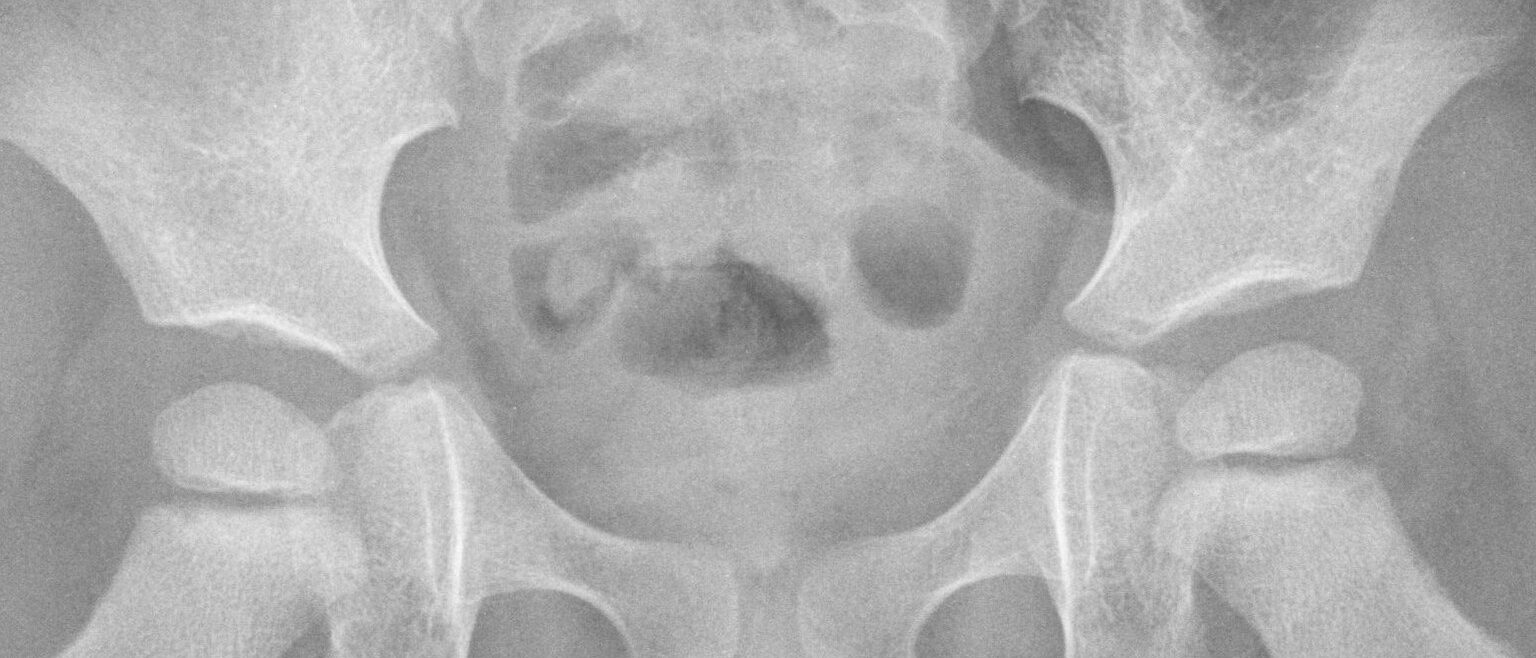
Who it is for
Senior trainees, fellows and early-career consultants who treat children's musculoskeletal conditions, worldwide.
The programme
25 one-hour sessions in 5 modules, each with its learning objectives. This is the draft programme: the course director reviews and refines it before enrolment opens, so session titles may change.
Module 1 · Sessions 1 to 5Assessing the child, infection, tumours and bone disease
1. Assessing the child: growth, normal variants and red flags
- Examine a child's gait, rotational profile and limb alignment and record the findings systematically
- Distinguish physiological in-toeing, bow legs, knock knees and flexible flat feet from pathology using age-specific norms
- Identify the red flags in history and examination that need investigation or referral
- Explain a normal-variant diagnosis to parents and agree a proportionate follow-up plan
2. The limping child and acute musculoskeletal infection
- Build an age-based differential diagnosis for the child with an acute limp
- Apply the BOAST on acute musculoskeletal infection in children to the investigation and timing of surgery
- Interpret clinical prediction rules (Kocher criteria) and recognise their limits in Kingella kingae infection
- Select empirical antibiotics, route and duration with the microbiology team and plan follow-up
3. Chronic infection and the inflammatory mimics
- Recognise subacute and chronic osteomyelitis on imaging and plan biopsy and culture
- Distinguish chronic non-bacterial osteomyelitis and juvenile idiopathic arthritis from infection
- Identify leukaemia and other malignancy presenting as limb or joint pain
- Decide when to refer to paediatric rheumatology or haematology and what to send with the referral
4. Bone tumours and tumour-like lesions in children
- Describe a bone lesion on plain radiographs using a structured approach and grade its aggressiveness
- Apply the NICE NG12 recommendation for very urgent X-ray in children with unexplained bone swelling or pain
- Manage common benign lesions: unicameral and aneurysmal bone cysts, non-ossifying fibroma, osteoid osteoma and osteochondroma
- Refer suspected sarcoma to a specialist centre before biopsy and explain why
5. Skeletal dysplasias and metabolic bone disease
- Recognise the clinical and radiographic features of common skeletal dysplasias and osteogenesis imperfecta
- Investigate and treat nutritional and hypophosphataemic rickets with the paediatric endocrinology team
- Plan fracture care and rodding in osteogenesis imperfecta alongside bisphosphonate treatment
- Outline the orthopaedic issues in achondroplasia and the status of new drug therapies such as vosoritide
Module 2 · Sessions 6 to 10The paediatric hip
6. Developmental dysplasia of the hip: screening and early treatment
- Describe the UK newborn and infant physical examination (NIPE) hip screening pathway and its risk factors, and the main US differences (AAOS guideline)
- Interpret hip ultrasound using the Graf method
- Manage a dislocatable or dysplastic hip with a Pavlik harness, including safe fitting and monitoring
- Recognise harness failure and avascular necrosis and decide when to stop and change treatment
7. Developmental dysplasia of the hip: late presentation and residual dysplasia
- Plan closed or open reduction with arthrogram and spica for a child who presents after harness age
- Select femoral shortening and pelvic osteotomy by age, acetabular index and remaining growth
- Classify avascular necrosis after DDH treatment and predict its effect on hip development
- Counsel families on the long-term outlook and the likelihood of further surgery
8. Perthes disease
- Stage Perthes disease and classify severity with the lateral pillar classification
- Use age at onset and hip containment to predict outcome
- Appraise the British Orthopaedic Surgery Surveillance (BOSS) study findings on containment surgery and on quality of life
- Agree a treatment plan with the family, including activity advice and when surgery is considered
9. Slipped capital femoral epiphysis
- Diagnose SCFE early, including in the child who presents with knee pain
- Classify slips by stability (Loder) and severity and explain the avascular necrosis risk of each
- Choose between in situ fixation and open reduction (modified Dunn, NICE IPG511) and decide on contralateral fixation
- Screen for endocrine and metabolic causes in atypical cases
10. The adolescent hip after childhood disease
- Assess hip pain in the adolescent after DDH, Perthes or SCFE
- Measure residual deformity and impingement on radiographs and cross-sectional imaging
- Select patients for periacetabular osteotomy, femoral osteotomy or impingement surgery
- Plan transition of hip care to adult services
Module 3 · Sessions 11 to 15The foot, the knee and lower limb deformity
11. Clubfoot
- Assess clubfoot severity with the Pirani score
- Describe the Ponseti method, including casting sequence and percutaneous tenotomy
- Explain foot abduction bracing to families and improve adherence
- Recognise and manage relapse, including repeat casting and tibialis anterior tendon transfer
12. The child's foot beyond clubfoot
- Distinguish flexible from rigid flat foot and identify tarsal coalition on imaging
- Recognise congenital vertical talus and plan its treatment
- Investigate the cavus foot for an underlying neurological cause such as Charcot-Marie-Tooth disease
- Manage idiopathic toe walking and decide when to investigate
13. Angular deformity and guided growth
- Analyse lower limb alignment with standing long-leg radiographs and the CORA method
- Distinguish physiological bowing from Blount disease and rickets
- Plan guided growth with tension band plates, including timing and rebound
- Decide when acute or gradual osteotomy is preferable to guided growth
14. Limb length discrepancy and congenital limb deficiency
- Predict limb length discrepancy at maturity with the multiplier method
- Choose between epiphysiodesis, lengthening and shoe raise according to predicted discrepancy
- Describe lengthening with external fixators and magnetic intramedullary nails, and their complications
- Outline staged reconstruction for fibular hemimelia, congenital femoral deficiency and congenital pseudarthrosis of the tibia
15. The paediatric and adolescent knee
- Manage juvenile osteochondritis dissecans according to lesion stability and skeletal maturity
- Recognise and treat symptomatic discoid meniscus
- Assess patellar instability in the growing child and choose growth-friendly stabilisation
- Apply the BOAST on ACL injury in the skeletally immature patient to the initial assessment and referral
Module 4 · Sessions 16 to 20Cerebral palsy and neuromuscular conditions
16. Cerebral palsy: assessment and the multidisciplinary team
- Classify function with the Gross Motor Function Classification System and relate it to orthopaedic goals
- Examine a child with cerebral palsy for spasticity, contracture and deformity
- Apply NICE NG62 to the orthopaedic and multidisciplinary care of children with cerebral palsy
- Select patients for botulinum toxin, serial casting and selective dorsal rhizotomy (NICE IPG373)
17. Hip surveillance and hip surgery in cerebral palsy
- Run a hip surveillance programme using migration percentage and GMFCS level
- Compare UK surveillance programmes (CPIP) with the AACPDM care pathway
- Select patients for soft-tissue release, femoral varus derotation and pelvic osteotomy
- Decide on salvage surgery for the painful dislocated hip and discuss goals with families
18. Gait analysis and single-event multilevel surgery
- Interpret instrumented gait analysis to identify the primary gait problems
- Recognise common gait patterns in diplegia and hemiplegia, including crouch and equinus
- Plan single-event multilevel surgery and the rehabilitation it needs
- Measure outcomes with validated functional scores
19. Neuromuscular conditions beyond cerebral palsy
- Apply the international Duchenne muscular dystrophy care considerations to contracture, fracture and spine management
- Plan orthopaedic care in spina bifida according to neurological level
- Manage foot and hip deformity in Charcot-Marie-Tooth disease and arthrogryposis
- Explain how disease-modifying therapies are changing the natural history in neuromuscular disease and what that means for surgical timing
20. Neuromuscular scoliosis and the medically complex child
- Assess the indications for spinal fusion in neuromuscular scoliosis
- Identify the cardiac, respiratory and nutritional risks that change perioperative planning
- Lead a goals-of-care discussion with families and the multidisciplinary team
- Describe the main complications and how to reduce them
Module 5 · Sessions 21 to 25Spine, upper limb and practice
21. Adolescent idiopathic scoliosis
- Assess the adolescent with scoliosis and measure the Cobb angle and skeletal maturity
- Predict curve progression and apply the BrAIST trial and SOSORT guidance to bracing decisions
- Explain the indications for spinal fusion and what families can expect
- Identify atypical features that need MRI
22. Early-onset scoliosis and back pain in children
- Classify early-onset scoliosis by cause and age
- Choose between Mehta casting, bracing and growth-friendly surgery
- Investigate the child with back pain and recognise red flags
- Manage spondylolysis and spondylolisthesis in young athletes
23. The paediatric upper limb
- Assess brachial plexus birth injury and decide when to refer for nerve surgery
- Manage shoulder deformity after brachial plexus birth injury
- Recognise Sprengel shoulder, congenital pseudarthrosis of the clavicle and congenital radial head dislocation
- Identify congenital hand differences that need early referral to a hand surgery service
24. Consent, safeguarding and transition
- Obtain valid consent from children, young people and those with parental responsibility, applying Gillick competence
- Act on safeguarding concerns that arise in elective clinics
- Resolve disagreements between families and the clinical team, including second opinions
- Plan transition of care to adult services for young people with long-term conditions
25. Paediatric orthopaedics worldwide and complex case decisions
- Adapt management of clubfoot, DDH and infection to settings with limited resources
- Manage neglected deformity and chronic osteomyelitis in children who present late
- Apply the course to complex cases in a panel discussion
- Build a personal plan for continuing development in paediatric orthopaedics
Fractures, non-accidental injury and polytrauma in children are taught in the Paediatric Trauma Diploma.
What the diploma includes
- 25 live sessions on Zoom, one evening a week, with recordings for the weeks you miss.
- Practising specialists teaching live, so you can put your questions to them.
- Weekly MCQs and a final written assessment.
- A digital certificate carrying your final classification.
Lead or teach the Paediatric Orthopaedics Diploma
We are looking for a course director and faculty from the specialty. Sessions are one hour in the UK evening, live on Zoom, and paid.